Picture Of Forearm Muscles And Tendons : Anatomy 101: Wrist Tendons - The Handcare Blog / Cross sectional anatomy of the upper limb :. The posterior compartment of the forearm (or extensor compartment) contains twelve muscles which are chiefly responsible for extension of the wrist and digits, and supination of the forearm. Antagonist of forearm flexors ( bra… flexion powerful of elbow and supination of forearm; The forearm has the shape of a somewhat flattened cone, being large above and small below. Also, pollicis means thumb in latin. The flexor compartment has three layers:
When identifying the function of the forearm muscles, it is important to note that any forearm compartment muscle that crosses the elbow joint will act at this joint. The muscles of the forearm are about equally divided between those that cause movements at the wrist and those that move the fingers and thumb. Know the causes, symptoms, treatment, recovery period and exercises for grade iii strain of forearm muscle: If you keep your hand flat on a table and. Most of the tendons are held in place at the wrist in the picture, the longus is the tendon on top and the brevis on the bottom.
Pictures Of Brachialis Tendons from healthiack.com The longer the muscles in the forearm are (and therefore the shorter their tendons are), the easier it will be to develop them. Anterior, lateral or posterior compartment. Do it yourself as shown in the picture! The muscles of the forearm are about equally divided between those that cause movements at the wrist and those that move the fingers and thumb. Find stockbilleder af forearm muscles tendons i hd og millionvis af andre royaltyfri stockbilleder, illustrationer og vektorer i shutterstocks samling. Supportive care for forearm muscle strain will involve following the rice protocol. It inserted independently into the. Each of these various compartments is.
The muscles of this group take origin from the medial epicondyle of the humerus by a common tendon;
The anterior forearm muscles are divided into 3 muscular layers ; All superficial muscles are arises from the medial epicondyle of humerus but they are inserted into the different part except. 12 (4 superficial + 3 mobile wad + 5 deep). Originates from the anterior surface of the ulna and attaches to the. Do it yourself as shown in the picture! The longer the muscles in the forearm are (and therefore the shorter their tendons are), the easier it will be to develop them. The pronator teres has two heads of. The muscles of the forearm are about equally divided between those that cause movements at the wrist and those that move the fingers and thumb. Anconeus muscle is a small muscle that is triangular in shape. Most of these originate from the lateral epicondyle. It originates from the lateral epicondyle of humerus via the common extensor tendon. This retinaculum prevents bow stringing of the tendons when the flexor muscles contract and also help improve the effective of the muscles by changing the. Find stockbilleder af forearm muscles tendons i hd og millionvis af andre royaltyfri stockbilleder, illustrationer og vektorer i shutterstocks samling.
Long flexor tendons extend from the forearm muscles through the wrist and attach to the small bones of the fingers and thumb. Tendons are under extreme stress when muscles pull on them, so they are very strong and are woven into the coverings of both muscles and bones. These types of strains are quite severe and involve complete rupture of the muscle fibers and tendons. Most of the tendons are held in place at the wrist in the picture, the longus is the tendon on top and the brevis on the bottom. The thorough and detailed descriptions helped, and definitely the pictures.
MS Review - Physical Therapy Pt101 with Review at Florida ... from classconnection.s3.amazonaws.com It inserted independently into the. All superficial muscles are arises from the medial epicondyle of humerus but they are inserted into the different part except. Superficial, intermediate two other structures of importance in the forearm are the common flexor tendon and the common extensor tendon. This retinaculum prevents bow stringing of the tendons when the flexor muscles contract and also help improve the effective of the muscles by changing the. Lesson on the anatomy of the forearm: It originates from the lateral epicondyle of humerus via the common extensor tendon. The posterior compartment of the forearm (or extensor compartment) contains twelve muscles which are chiefly responsible for extension of the wrist and digits, and supination of the forearm. 12 (4 superficial + 3 mobile wad + 5 deep).
If you keep your hand flat on a table and.
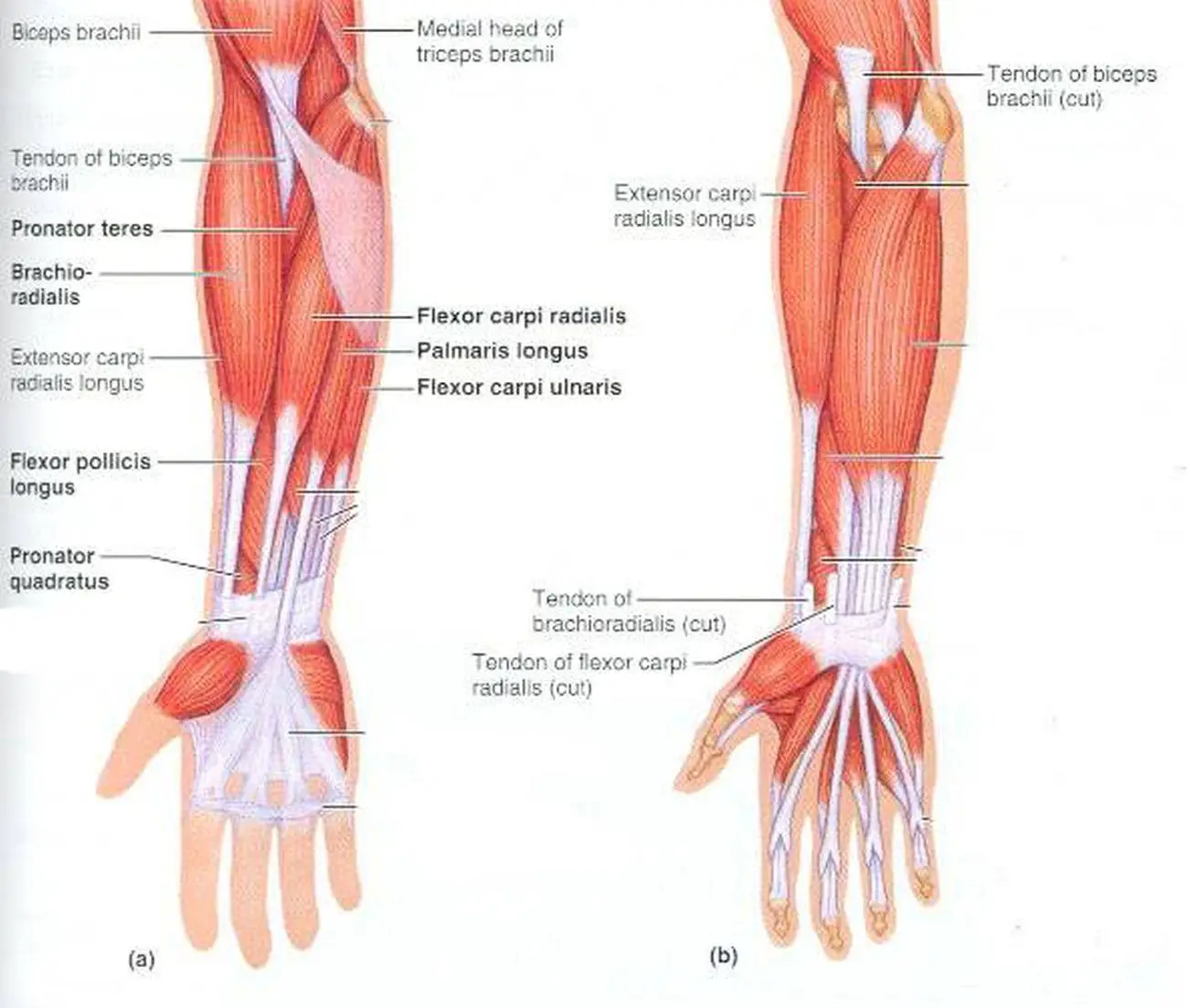
The posterior compartment of the forearm (or extensor compartment) contains twelve muscles which are chiefly responsible for extension of the wrist and digits, and supination of the forearm. It originates from the lateral epicondyle of humerus via the common extensor tendon. If you keep your hand flat on a table and. The muscles of the forearm are numerous, differ in the variety of functions. Forearm muscles are usually divided into an anterior flexor compartment and a posterior extensor compartment. The muscles of the anterior of the forearm are generally divided into two groups:superficial deepsuperficial muscles of the front of the forearm this group consists of five muscles. See anatomy pictures of the 27 bones in the hand and wrist, how they are connected with tendons and muscles and the nerves that run through the skeletal structure. The forearm has the shape of a somewhat flattened cone, being large above and small below. The muscle fibers then descend towards the wrist area where they converge onto a narrow tendon. 12 (4 superficial + 3 mobile wad + 5 deep). All 4 muscles have a common origin at the medial epicondyle of the humerus, known as the common flexor tendon. Originates from the anterior surface of the ulna and attaches to the. The muscles of the forearm are predominantly slow twitch. slow twitch muscles are very resistant alternate days so that the muscles and tendons have time to recover from the previous workout.
The forearm is the region of the upper limb between the elbow and the wrist. Originates from the anterior surface of the ulna and attaches to the. Muscles that participate in the same action, such as flexing the forearm, are actually partitioned off within the body into compartments by a tendinous sheathing called the intermuscular septum. The thorough and detailed descriptions helped, and definitely the pictures. The posterior compartment of the forearm (or extensor compartment) contains twelve muscles which are chiefly responsible for extension of the wrist and digits, and supination of the forearm.
Forearm Muscles - Structure, Injuries, Veins & Exercise ... from www.muscleseek.com All 4 muscles have a common origin at the medial epicondyle of the humerus, known as the common flexor tendon. The muscles of this group take origin from the medial epicondyle of the humerus by a common tendon; The extensor carpi ulnaris muscle is the most medial muscle in the superficial posterior compartment of the forearm. Originates from the anterior surface of the ulna and attaches to the. Tendons are under extreme stress when muscles pull on them, so they are very strong and are woven into the coverings of both muscles and bones. The forearm has the shape of a somewhat flattened cone, being large above and small below. Hold your elbow with thumbs up and other 4 extension of index finger. Antagonist of forearm flexors ( bra… flexion powerful of elbow and supination of forearm;
Arm muscles can also be classified by their compartments or regions.
Forearm muscles are usually divided into an anterior flexor compartment and a posterior extensor compartment. There are many muscles in the forearm. Hold your elbow with thumbs up and other 4 extension of index finger. This is because the bellies of the muscles lie above and their 0shares facebook twitter reddit flipboard linkedinwelcome back to the series that loves to talk about muscle, and is therefore aptly named. One of these originated from the extensor carpi radialis brevis, became tendinous and travelled between the two radial extensor tendons. The muscles of the forearm are predominantly slow twitch. slow twitch muscles are very resistant alternate days so that the muscles and tendons have time to recover from the previous workout. It inserted independently into the. Most of these originate from the lateral epicondyle. The muscle fibers then descend towards the wrist area where they converge onto a narrow tendon. They control movements of the wrist, hand, fingers and thumb. Most of the muscles are multiarticular at the level of the middle of the forearm, the muscular abdomen continues into a narrow flat tendon that passes under the tendons of the long distal muscle and the short extensor of. Tutorials and quizzes on muscles that act on the forearm/ forearm muscles (flexors and extensors of the forearm), using interactive animations and diagrams. Antagonist of forearm flexors ( bra… flexion powerful of elbow and supination of forearm;
Long flexor tendons extend from the forearm muscles through the wrist and attach to the small bones of the fingers and thumb picture of forearm tendons. Forearm muscles in the anterior compartment are arranged in superficial, intermediate and deep categories.


Posting Komentar
0 Komentar